My Favorite Movie Quote -
Yeah, but your scientists were so preoccupied with whether or not they could, they didn't stop to think if they should.
- Dr. Ian Malcolm - Jurrasic Park

Monday, February 28, 2011

Feb 28, 2011 - Skull Features and Personality

http://phillips.blogs.com/photos/uncategorized/12phrenology.jpg
  
Phrenology is the science which studies the relationships between a person's character and the morphology of the skull. It is a very ancient object of study. The first philosopher to attempt to (my words) locate mental faculties in the head was in fact Aristoteles.  http://www.phrenology.org/intro.html

In modern terms Phrenology is a spurious method for assessing aspects of character and personality from the physical appearances of certain areas of the cranium. Like physiognomy in general, there was a time when the issues of phrenology were presented as empirical or scientific questions, but as the preposterous claims of practitioners became more obviously bogus, this field was classified with the pseudo-sciences by the end of the 19th century. In the 20th century, these topics have increasingly been associated with the mysterious or occult literature, and have acquired the stigma of this association. Perhaps this shift is an aspect of continuing to market books and other products covering this material.  http://face-and-emotion.com/dataface/physiognomy/phrenology.jsp


In the example from Lehmann below, each numbered area is relevant to a personal characteristic. For example, area 20 is related to how confident or imbued with faith one is. If area 20 is enlarged, the person is confident, but if it is lacking, the person is critical, not confident. Area 27 has to do with the sense of humor. If it is enlarged, the person is easily drawn into laughter, but if it is lacking, the person is too serious to appreciate jokes.

From: Arthur R. H. Lehmann. Krankheit Begabung Verßprechen. Berlin: F. Gnadenfeld co., 1904.

Some phrenologists tried to supply answers to deeper questions and to connect the issues of phrenology to those of physiognomy. Spurzheim saw the angles of the face as an important feature of the face, and he knew that the bony structure of the skull was the basis for these angles and other features. Using such observations, he claimed that four personality types were connected physiologically to how the skull was shaped. The validity of such connections is doubtful, but many subsequent researchers have tried to show how body shapes and sizes are related to personality variables, siting more evidence to support their contention. In general, however, the relationships between body characters, including those of the face and head, and psychological variables have not been systematically compiled and evaluated, and the influence of such approaches has been minimal.

The term "physiognomy" refers to features of the face, especially so skull - basis of physiognomy 
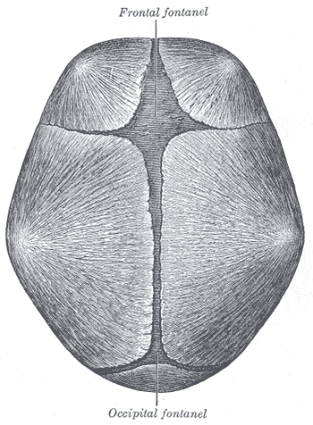
when, in the narrow sense, these features are used to infer the relatively enduring character or temperament of an individual. In this discussion, physiognomy connotes a broader meaning, i.e., it refers to relatively unchanging facial features that might convey messages about any inner or hidden aspect of the person. Most of these facial features have as their basis the bony structure of the skull, on which the soft tissues lie. These features include the shapes and positions of major areas and landmarks of the face, such as the forehead, eyebrows, nose, cheeks, and mouth. The important facial features can be fairly accurately reconstructed by experts from the skull alone. A diagram of the human cranium shows the major features of the skull, from which much of the visible appearances of the face can be extrapolated. Other physiognomic features are not directly linked to the bony skull, such as skin texture and coloration, hair placement and texture, and detailed shapes of fleshy features. All of these features change slowly and relatively little over time, and they are the sign vehicles for physiognomic messages. Proposing an association between these facial features and other aspects of the person, including personality, character, outcomes of medical treatment, romantic compatibility,or the destiny of the person, is a physiognomic approach. The validity of the association or inference based on physiognomy is a separate issue that can be established or discredited by empirical evidence. Accurately face reading these signs depends upon knowing which relations are valid and which are spurious.  For more information see the following link http://face-and-emotion.com/dataface/physiognomy/physiognomy.jsp

Fun facts:
- In Bram Stoker's Dracula several characters make phrenological observations in describing other characters.

- Terry Prachett, in his Discworld series of books, describes the practice of Retro-phrenology as the practice of altering someone's character by giving them bumps on the head. You can go into a shop in Ankh-Morpork (a city in the book) and order an artistic temperament with a tendency to introspection. What you actually get is hit on the head with a series of small hammers, but it keeps the money in circulation and gives people something to do.

Friday, February 18, 2011

Feb 18, 2011 Fontanelles & Joining the Cranial Bones

The skull is made up of many bones, 7 in the skull itself and 14 in the facial area. They join together to form a solid, bony cavity that protects the brain and supports the structures of the head. The areas where the bones join together are called the sutures.

The bones are not joined together firmly at birth. This allows the head to pass through the birth canal. The sutures get minerals added to them over time and harden, firmly joining the skull bones together. This process is called ossification.


In an infant, the space where two sutures join forms a membrane-covered "soft spot" called a fontanelle (fontanel). The fontanelles allow for growth of the skull during an infant's first year.
There are normally several fontanelles on a newborn's skull, mainly at the top, back, and sides of the head. Like the sutures, fontanelles ossify over time and become closed, solid bony areas. The fontanelle in the back of the head (posterior fontanelle) usually closes by the time an infant is 1 - 2 months old. The fontanelle at the top of the head (anterior fontanelle) usually closes between 7 - 19 months.
The fontanelles should feel firm and very slightly curved inward to the touch. A tense or bulging fontanelle occurs when fluid builds up in the skull cavity or when pressure increases in the brain (increased intracranial pressure).


      http://en.wikipedia.org/wiki/File:Gray198.png


When the infant is crying, lying down, or vomiting, the fontanelles may look like they are bulging, but they should return to normal when the infant is in a calm, head-up position.
http://www.nlm.nih.gov/medlineplus/ency/article/003310.htm


Many animals are born with fonatanelles. These normally close at various points during maturation.  During the intervening time the brain is not as well protected as it will be and care should be taken when handling very young animals.  Some animals, such as dogs, will have fontanelles that don't close.  Which can cause difficulties for the dog (and the dog owner).  Pressure on the soft spot can cause brain damage and seizures.
                 
Fun fact -  There is no law at the U.S. Federal level prohibiting you from having a human bone in your possession. The fact that some people believe there is or believe there should be such a law is irrelevant. 
For more information go to  http://www.boneroom.com/faqs/bones.html#bonelaws

Thursday, February 17, 2011

Feb 17, 2011 Where Man Meets Machine

There seems to be an inborn need in many humans to create things in our likeness.  Novelists and movie directors have taken this endeavor to the extreme in writing stories and creating excellent horror, action, and mystery movies to intrigue us.  Asimov's i,Robot  series (later turned into a movie starring Will Smith) blurs the line between robot and human. 


The Terminator series takes us into the future and back to the present as Arnold Schwarzenegger (along with other actors play robots that are  trying to prevent humanity from defeating them in the future.  By destroying the humans from the past that will eventually defeat them. 


Robin William's take on AI (artificial intelligence) in Bicentennial Man and Star Trek's Data, the android played by Brent Spiner (who does a wonderful job portraying Data's fledgling emotional growth), take a more benign look at the interaction between humans and AI.


These portrayals have always provided us with great entertainment, but as we merge our continuing studies of Anatomy, Physiology, and Neurology with robotics, computer programing, and artificial intelligence we are starting to approach something that might make us consider where this is all headed.


These are just two of a large number of videos, quite easily found online, that indicate the direction we are moving in; but why?  There are actually a multitude of applications (far too many to be listed here) ranging from the scary science-fiction-like the Clone Wars' militaristic application to a butler-like human aide as represented in the movie classic Star Wars by C3PO.

If you pay attention to the majority of the stories in which the Robot/Android plays a major role I think you will often find that most of them play out as a look into a long debated question.  

"What is life?"

When does AI cross over from - I think because you ask it of me, to - I think, therefore I am?


Fun fact - Long before Isaac Asimov first touched a typewriter, Walt Whitman's boon companion William Douglas O'Connor toiled over a story of steampunk robots and political intrigue, called "The Brazen Android."

A list of worthwhile Robot/Android stories (some of which are now movies):
  • Do Androids Dream of Electric Sheep? (Now called Blade Runner)
  • The Terminator Series
  • Bicentennial Man
  • The i,Robot series by Asimov
  • some specific episodes of Star Trek - Next Generation

Please add your favorites in the comment section.

Tuesday, February 15, 2011

Feb 15, 2011 - The Axial & Appendicular Skeleton

Reminder - Thursday 2/17 is the bone Histology Quiz

The Axial and Appendicular Skeleton together cover the following functions:

  1. STRENGTH, SUPPORT AND SHAPE: It gives strength, support and shape to the body. Without a hard and rigid skeletal system, human body cannot stand upright, and it will become just a bag of soft tissues without any proper shape
  2. PROTECTION OF DELICATE ORGANS: In areas like the rib cage and skull, the skeleton protects inner soft but vital organs like heart and brain from external shocks. Any damage to these organs can prove fatal, therefore protective function of skeleton is very important
  3. LEVERAGE FOR MOVEMENTS: Bones of the human skeleton in all parts of body provide attachment to the muscles. These muscles provide motor power for producing movements of body parts. In these movements the parts of skeleton acts like levers of different types thus producing movements according to the needs of the human body.
  4. PRODUCTION OF RED BLOOD CELLS: Bones like the sternum, and heads of tibia have hemopoeitic activity (blood cells production). These are the sites of production of new blood cells.
The Axial Skeleton

The axial skeleton:
  • is made of the skull, spine, sacrum, and rib cage 
  • allows various movements of the torso at the vertebrae but it is primarily for protection 
  • has  80 of the 206 bones of the body
The Appendicular Skeleton


http://www.mananatomy.com/wp-content/uploads/2010/12/appendicular-skeleton.jpg
The appendicular skeleton:
  • is made of the shoulder girdle, upper limbs (appendages), pelvic girdle,  and lower limbs
  • is made primarily for leverage/movement/locomotion
  • has  126 of the 206 bones of the body
Fun fact:  A popular theory regarding skeleton bones is that men have one less bone than women, owing to the Bible story that Eve was created from one of Adam's ribs. Both men and women actually possess 12 pairs of ribs. It is interesting to note; however, that should a rib need to be removed, it can regenerate itself within 3 months.

    Monday, February 14, 2011

    Feb 14, 2011 Happy Valentine's Day or Black Monday (Whichever fits)

    Reminder - Thursday 2/17 is the bone Histology Quiz


    Today is a departure from strict anatomy. Many students have asked me why we draw a heart the way we do today.  Here is a little information.  I will not venture to say which if any of these are correct.  See the following link for more information. http://www.romancestuck.com/articles/general/history-of-heart-shape.htm

    In modern days, the heart symbolizes love, romance, and passion, but where did this come from?

    The History of the Heart Shape

    The Heart Shape
    The heart is traditionally drawn in this shape with a red color.
    Colors of the Heart
    The color of hearts is generally red colored. Red traditionally represents passion and strong emotions in many cultures.

    Heart Theories
    From what we could determine, there are three main theories as to where the heart shape originated. None of the three are proven, but all present interesting points:  

    1. Modeled after an actual heart.
    This theory is actually a pretty well known one. The theory says that the modern drawing of a heart is actually modeled after the actual human heart because the ancients, including Aristotle, and even some today, believe that the heart contains all human passions. However, the traditional heart shape as drawn has very little resemblance to an actual human heart. The drawing more closely matches that of a cattle heart, which was more readily available in sight in past centuries, meaning they modeled it after a cattle heart. However, the resemblance to the cattle heart is still very slight. It's also said that the drawing of a modern heart possibly originated because of botched drawings of the human heart by medieval artists after a philosopher inaccurately described the shape. The medieval tradition of courtly love may have reinforced the shape's association with romance. Hearts can be found on playing cards, tapestries, and paintings.
     Human heart - The most "heart shaped" I could find.

    2. Depicts the female body.
    Some say that the traditional heart shape actually represents body features of a female, such as the vulva, breasts, or buttocks. The female pelvis is also somewhat heart-shaped. Because the female gives birth to life, this could be the association with the heart-shape and love.

    3. Derived from a seed.
    A last theory is that the traditional heart came was derived from the plant seed of the Silphium plant. This now extinct (for over two millennia) North African plant was found in the ancient city of Cyrene during the seventh century BC. The plant was most commonly used as a seasoning, but was also widely used as a contraceptive in ancient Egypt. Because of it's importance to their economy, they depicted the shape of the seed on their coins. The seeds and flowers were distinctly heart-shaped. It is said that because of the use of the plant as a contraceptive, the shape of the seeds became associated with sexuality and eventually with romantic love.

    Silphium Seed Coin - http://growingupcreative.typepad.com/.a/6a010535781c42970c011278d8daef28a4-pi

    Fun Fact -  One of the most ancient of Valentine's Days rituals (dating from at least the Middle Ages and possibly earlier) was the practice of writing the names of young ladies on slips of paper and placing them within a jar or bowl. The lady whose name was drawn by an eligible bachelor became his valentine and he wore the name on his sleeve for one week. It is believed that the saying "to wear one's heart on one's sleeve" (meaning that is is easy for others to know the romantic inclination of an individual) may have originated from this custom.

    Valentine Superstition - It was believed that if a lady slept with a sprig of rosemary pinned inside the pillow on the Eve of Valentine's Day it would encourage dreams of a future sweetheart's face.

    For more information try the following - http://www.novareinna.com/festive/valrites.html

    Friday, February 11, 2011

    Feb 11, 2011 Unbreakable vs. Osteogenesis Imperfecta


         Elijah Price (Samuel L. Jackson) is born with Type I osteogenesis imperfecta, a rare disease in which bones break easily.  He grew up with the nickname "Mr. Glass."  Drawing on what he has read in comic books during his many hospital stays, Price theorizes that if he is frail to the extreme, then perhaps there is someone strong to the opposite extreme.
         David Dunn (Bruce Willis) is Price's polar opposite which he discovers slowly throughout the film as Price tries to convince him that he is the mordern incarnation of a superhero.  Dunn eventually begins to believe it himself and after discovering he has a limited form of ESP begins to embody the idea of a minor hero.  He also finds that he has a weakness that nearly kills him.  By the end of the film Dunn confronts Price about his involvement in finding someone (Dunn) who is his archnemesis.

        The purpose of this blog is not to review a movie but to take a brief look at the disease osteogenesis imperfecta.

    Osteogenesis Imperfecta

    Osteogenesis imperfecta (OI) is a congenital disease, meaning it is present at birth. It is frequently caused by defect in the gene that produces type 1 collagen, an important building block of bone. There are many different defects that can affect this gene. The severity of OI depends on the specific gene defect.
    OI is an autosomal dominant disease. That means if you have one copy of the gene, you will have the disease. Most cases of OI are inherited from a parent, although some cases are the result of new genetic mutations.

    All people with OI have weak bones, which makes them susceptible to fractures. Persons with OI are usually below average height (short stature). However, the severity of the disease varies greatly.
    The classic symptoms include:
    Because type I collagen is also found in ligaments, persons with OI often have loose joints (hypermobility) and flat feet. Some types of OI also lead to the development of poor teeth.
    Symptoms of more severe forms of OI may include:

    Treatment

    There is not yet a cure for this disease. However, specific therapies can reduce the pain and complications associated with OI.  In severe cases, surgery to place metal rods into the long bones of the legs may be considered to strength the bone and reduce the risk of fracture. Bracing can also be helpful for some people.

    Fun Fact: In the movie GATTACA Vincent has this procedure done to increase his height.  He is attempting to impersonate someone else.  Consider what he is dong in light of the quotes below presented at the very beginning of the movie.

    "Consider God's handiwork; who can straighten what He hath made crooked?" - Ecclesiastes 7:13

    "I not only think that we will tamper with Mother Nature, I think Mother wants us to." - Willard Gaylin

    Thursday, February 10, 2011

    Feb 10, 2011 - The Long & Short of Bone Growth

    Long Bone Growth

    Long bones add materials at specific areas called growth plate. The growth plate, also known as the epiphyseal plate or physis, is the area of growing tissue called hyaline cartilage near the ends of the long bones in children and adolescents. Each long bone has at least two growth plates: one at each end. The growth plate determines the future length and shape of the mature bone. When growth is complete - sometime during adolescence - the growth plates close and are replaced by solid bone. In adults, who have stopped growing, the plate is replaced by an epiphyseal line. Because the growth plates are the weakest areas of the growing skeleton - even weaker than the nearby ligaments and tendons that connect bones to other bones and muscles - they are vulnerable to injury. Injuries to the growth plate are called fractures.

    This is the distal end of the tibia (left) and fibula (right).  
    The thin lines most proximal on both bones are the epiphyseal (growth) plates.
    http://upload.wikimedia.org/wikipedia/en/7/7a/Tib_fib_growth_plates.jpg

    About 85 percent of growth plate fractures heal without any lasting effect. Whether an arrest of growth occurs depends on the treatment provided, and the following factors, in descending order of importance:
    • Severity of the injury - If the injury causes the blood supply to the epiphysis to be cut off, growth can be stunted. If the growth plate is shifted, shattered, or crushed, the growth plate may close prematurely, forming a bony bridge or “bar.” The risk of growth arrest is higher in this setting. An open injury in which the skin is broken carries the risk of infection, which could destroy the growth plate.
    • Age of the child - In a younger child, the bones have a great deal of growing to do; therefore, growth arrest can be more serious, and closer surveillance is needed. It is also true, however, that younger bones have a greater ability to heal.
    • Which growth plate is injured - Some growth plates, such as those in the region of the knee, are more involved in extensive bone growth than others.
    • Type of fracture
    The most frequent complication of a growth plate fracture is premature arrest of bone growth. The affected bone grows less than it would have without the injury, and the resulting limb could be shorter than the opposite, uninjured limb. If only part of the growth plate is injured, growth may be lopsided and the limb may become crooked.
    Growth plate injuries at the knee have the greatest risk of complications. Nerve and blood vessel damage occurs most frequently there. Injuries to the knee have a much higher incidence of premature growth arrest and crooked growth.   http://www.niams.nih.gov/Health_Info/Growth_Plate_Injuries/default.asp#8
          
         Using the principal that bone will grow (relatively) rapidly to fill in areas of fracture Ilizarov discovered that by carefully severing a bone without severing the periosteum around it, one could separate two halves of a bone slightly and fix them in place, and the bone would grow to fill the gap. He also discovered that bone regrows at a fairly uniform rate across people and circumstances.
         These experiments led to the design of what is known as an Ilizarov apparatus, which holds a severed bone in place, by virtue of a framework and pins through the bone, and separates halves of the bone by a tiny amount; by repeating this over time, at the rate of the bone's regrowth, it is possible to extend a bone by a desired amount.
    http://en.wikipedia.org/wiki/Ilizarov_apparatus
    Ilizarov Apparatus

    • Bone infections
    • Poliomyelitis Sequelae (limb lengthening and correction of deformities)
    • Treatment of non-unions and malunited fractures
    • Correction of deformities, both congenital & acquired, of the limbs 
    • Badly comminuted fractures (multiple fragments) in the limbs, even with skin loss.
    This procedure is accepted for the conditions listed above but is very controversial for cosmetic limb lengthening.  The article at the following link provides multiple viewpoints on the procedure.  It is very interesting.   http://www.shortsupport.org/News/0432.html 

    Fun Fact:  I the movie GATTACA the character Vincent has this procedure done as he gets ready to take the identity of Jerome.  This is part of the plot that follows the following Title Cards:

    [first title card]
    "Consider God's handiwork; who can straighten what He hath made crooked?" - Ecclesiastes 7:13

    [second title card]
    "I not only think that we will tamper with Mother Nature, I think Mother wants us to." - Willard Gaylin

    Wednesday, February 9, 2011

    Feb 8, 2011 - Throw me a Bone Here! - Extra Bones?

    Reminder - The Histology Comprehensive Test is this Friday 2/11. It will be run as a practical exam.

    Wormian Bones & Accessory Bones

    http://upload.wikimedia.org/wikipedia/commons/9/93/Wormian_bones.jpg

    Wormian bones are a subset of the small intrasutural bones that lie between the cranial sutures formed by the bones of the skull vault. The title Wormian bones is given to abnormal intrasutural bones that are typically found around the lambdoid suture.
    Erroneously, these bones are named after the person who was supposed to have first described them:
    • Ole Worm (1624-1639): professor of Anatomy at Copenhagen, Denmark
    http://radiopaedia.org/articles/wormian-bones

    Accessory Bones

    The image above shows a common form of often problematic accessory bone.

         These are "extra" bones resulting, most often, from altered patterns of ossification. The accessory bones (also called ossicles) occur most often in the hands, wrists and feet. Examples include the os naviculare secundarium, carpal boss, os trigonum, os vesalianum, os intermetatarsale, and os supratrochleare dorsale. These ossicles are generally not considered significant and are believed to represent variations of normal development. It is not uncommon for both the wrists and/or the feet to contain extra small ossicles, that occasionally cause pain. It is important to note that other problems such as avulsions or chip fractures may sometime be misidentified as accessory bones.    http://www.medcyclopaedia.com/library/topics/volume_iii_1/a/accessory_bones.aspx

     Fun Fact: The soft spot(s) on a baby's head called fontanelles are the places that Wormian bones most often
                    form due to the pattern of ossification in the baby's head.

    Tuesday, February 8, 2011

    Feb 8, 2011 - Bone Construction


    Reminder - The Histology Comprehensive Test is this Friday 2/11.
                      It will be run as a practical exam.
                   - Check out this website for reviewing for the test, it is the best one yet
                      http://nhscience.lonestar.edu/biol/tissue.html

    Anatomy of a Bone

    http://www.baileybio.com/plogger/images/biology/powerpoint_-_integumentary__skeletal__and_muscular_systems/bone_tissue.jpg

         The image above gives a good overview of the gross anatomy and the micro-anatomy of a long bone.  You should become familiar with the definitions of these terms as well as be able to identify them on images in a  quiz setting.
         Bone tissue is "living"  in that it is constantly changing and is very responsive.  The bone material will change its shape to meet the demands of forces put on it.  
         The image below shows the result of Chinese foot binding.  The bones in the foot change shape in response to the external pressures exerted on them.

    Foot Binding (纏足)


    In the 10th   century, in China, legend says a prince began the practice of foot binding because he loved the small "Lily Feet" of his concubine. The tiny foot became the mark of a wealthy and well-born woman.
    Footbinding was a custom practised on young females for approximately one thousand years in China, and ended in the early 20th century.  In Chinese foot binding, young girls’ feet, usually at age 6 but often earlier, were wrapped in tight bandages so that they could not grow and develop normally; they would, instead, break and become highly deformed, not growing past 4-6 inches (10-15 cm).  Bound feet had to be washed and cared for daily.  If toe nails grew into the instep, infection could set in.  If the bindings were too tight, gangrene and blood poisoning could occur.  The bound foot was painful and tender forever.  It often had an unpleasant smell.  Today, the result of foot binding is a prominent cause of disability among some elderly Chinese women.

    Plagiocephaly


    http://bloximages.chicago2.vip.townnews.com/heraldextra.com/content/tncms/assets/editorial/e/cd/da4/ecdda4ec-e86c-514f-8b87-b838075143b7.image.jpg

    What is Plagiocephaly? - Plagiocephaly occurs when a baby's head develops a flat spot or becomes asymmetrical due to some kind of external pressure. Many babies are born with an abnormally shaped head as a result of the pressure exerted on them during birth, but most babies' heads will correct themselves within about six weeks.  Plagiocephaly can also occur after childbirth and is not uncommon in neglected infants who are left to lie unattended for long periods.  The weight of the baby's head presses down on the back of the skull and the bones respond by forming to that shape.  http://www.babycenter.com/0_plagiocephaly-flat-head-syndrome_1187981.bc


    http://t2.gstatic.com/images?q=tbn:ANd9GcQIdBEyEIx6kda4AO790xIWbQVVgScdjSkXevDDHluYilYzHhwT
    Fun fact:   The crystal skulls (upon which the Indiana Jones movie are based) are a number of human skull hardstone carvings made of clear or milky quartz rock, known in art history as "rock crystal", claimed to be pre-Columbian Mesoamerican artifacts by their alleged finders. However, none of the specimens made available for scientific study have been authenticated as pre-Columbian in origin. The results of these studies demonstrated that those examined were manufactured in the mid-19th century or later, almost certainly in Europe.

    Monday, February 7, 2011

    Feb 7, 2011 - Boning Up

    Welcome back from the weekend.  I hope you had fun.  I spent 6 hours shoveling my roof and scraping my driveway. WooHoo!!!

    Reminder - The Histology Comprehensive Test is this Friday 2/11. It will be run as a practical exam.


    The following unit will deal with the skeletal system.  We will be examining the skeletal system from the micro-anatomy to the gross anatomy as well as studying how it grows and repairs itself.  We will also do some comparative anatomy and finally discuss the affects of aging and disease on the skeletal system.

    Fun Fact - Our bones are six times stronger than steel if both are the same weight.


    Friday, February 4, 2011

    Feb 4, 2011 - Cardiac Muscle Tissue

    Reminders:  
    • The C.T. Quiz has been moved to Mon 2/7.
    • The due date for the Histology project is now Mon 2/7.
    • The Muscle/Nervous Tissue Quiz will now be Wed 2/10.

      When trying to identify cardiac muscle tissue there are some easy markers to look for to help you
      make your choice.
        • The most important marker is the presence of intercalated disks which are special extensions of the cardiac cell membrane that has many functions.
        • The cells are often branched.
        • Each cell has only one nucleus but it may not always be visible. (It depends on the depth of the section taken through the cell.)
    Fun fact:   Chocolate can actually improve cardiovascular health, according to the Northwest Health Sciences University.  Chocolate contains cocoa which has antioxidants that help protect the cardiovascular system. The antioxidants help lower blood pressure and help reduce deaths related to cardiovascular disease. A 4-5 gram (less than an ounce) serving per day is enough to gain the benefits from the antioxidants found in cocoa. (If this is the case my heart may never die!)

    Read more: Fun Facts of the Cardiovascular System | eHow.com http://www.ehow.com/about_5079091_fun-cardiovascular-system.html#ixzz1Cvnv4P8D

    Thursday, February 3, 2011

    Feb 3, 2011 - C.T. Quiz Primer

    Reminders:  (Ok I lied we have moved the dates.  See below.)
    • The C.T. Quiz has been moved to Mon 2/7.
    • The due date for the Histology project is now Mon 2/7.
    • The Muscle/Nervous Tissue Quiz will now be Wed 2/10.
    (Click the images to enlarge them.)

      The drawings above give a quick overview of Connective Tissues using an artist's representation.  The fiber types and cell types are well represented.  Make sure to review Table 5.4 to help prepare for the upcoming quiz.

      The image below provides some microscope images of many of the same tissues.

      http://www.nicerweb.com/doc/class/bio1151/Locked/media/ch40/40_05ConnectStructFunc.jpg

      I have included some links below to help you practice for the upcoming quiz.

      http://www.unomaha.edu/hpa/2740connectivetissue.html#dense


      https://staff.rockwood.k12.mo.us/fullertonheather/hap/Documents/Unit%204%20Tissues%20and%20Integumentary%20System/Connective%20tissue%20practice%20test.ppt

      http://faculty.sdmiramar.edu/KPETTI/Bio160/MiscImages/ConnTissuChrt.jpg

      Fun Fact - In general, an excess of 3500 Calories will create 1 lb of body fat.  So removing 500 Calories a day whether by diet, exercise or both combined will allow the average person to drop 1 lb a week.  That doesn't sound like much until you realize that would be 52 lbs in one year!

      Wednesday, February 2, 2011

      Feb 2, 2011 - Skeletal Muscle

      Reminders: (None of these dates will change due to the weather!)
      1. Don't forget your connective tissue quiz is on Friday 2/4.
      2. The Histology Project is due Friday 2/4.
      3. Muscle & Nervous tissue quiz next Tuesday 2/8.
      4. Due to the many weather related issues we have had you may use the internet to research two of your histology drawings. Please include the following on your poster:
          1. The url as a reference
          2. The magnification (if possible)
      Skeletal Muscle Tissue
                                                              http://clcpages.clcillinois.edu/home/bio567/pages/newtissues/skeletalmuscle.htm

      Pictured above is a good example of skeletal muscle tissue.  It is important to note a few things about this tissue when trying to identify it:
      • the cells are generally parallel and unbranched (distinguishes it from cardiac or smooth muscle tissue)
      • each cell is multinucleated (distinguishes it from cardiac or smooth muscle tissue)
      • the cells have striations (stripes) running perpendicular to the length of the cell (distinguishes it from smooth muscle tissue)
      In a later unit we will investigate: some of the myths surrounding muscle/cell growth, how these cells produce force, what allows us to specify the strength of a muscle contraction, how training specificity changes a muscle, red vs. white fibers, and many more topics.

      If you have any questions regarding muscle anatomy for our class in the future please post them in the comments section.

      Fun Fact- It is a total myth (but commonly believed) that you can turn fat into muscle with training.

      Tuesday, February 1, 2011

      Jan 31, 2011 - Up and Running!

      Welcome to my first blog - of any sort, ever! 

      I am starting this blog to communicate with my Anatomy and Physiology students, Trimester 2, 2011. I will be posting a variety of materials here including: pictures, diagrams, informational links, practice tools and games, assignment updates, snow event information, a body-part of the day, and possible addendums to class materials and discussions.  I will check the comments often with the last check at around 8:30 in the evening (whenever possible). 
      I will try to post daily (but not often on weekends).  I expect to set the Blog up with some enrichment/extra credit assignments that expand upon the current information.  I'm sure I will run into snags and consider differnt formats and ways to use this as time goes on.
      I look forward to hearing from you and would appreciate suggestions for making this more useful as well as constructive criticisms of the posts as things progress.

      Fun Fact - An average human scalp has 100,000 hairs. Mr. Bernier's scalp has far less than that.

      Feb 1, 2011 - Blood

      Reminders:
      • Histology Projects are due Friday, 2/4 
      • Connective Tissue Quiz will be the same day.
      Part of the day - Blood
      Blood is considered a connective tissue for two basic reasons: (1) embryologically, it has the same origin (mesodermal) as do the other connective tissue types and (2) blood connects the body systems together bringing the needed oxygen, nutrients, hormones and other signaling molecules, and removing the wastes. In circulating blood two different cell types are found: enucleated erythrocytes or red blood cells and nucleated leukocytes or white blood cells. (http://lifesci.rutgers.edu/~babiarz/bloodtx.htm)


       Note - the part of the blood that is missing  (Difficult to do since it is not labeled!) the plasma.

      Plasma is the liquid component of blood, in which the red blood cells, white blood cells, and platelets are suspended. It constitutes more than half of the blood's volume and consists mostly of water that contains dissolved salts (electrolytes) and proteins. The major protein in plasma is albumin. Albumin helps keep fluid from leaking out of blood vessels and into tissues, and albumin binds to and carries substances such as hormones and certain drugs. Other proteins in plasma include antibodies (immunoglobulins), which actively defend the body against viruses, bacteria, fungi, and cancer cells, and clotting factors, which control bleeding.

      Plasma has other functions. It acts as a reservoir that can either replenish insufficient water or absorb excess water from tissues. When body tissues need additional liquid, water from plasma is the first resource to meet that need. Plasma also prevents blood vessels from collapsing and clogging and helps maintain blood pressure and circulation throughout the body simply by filling blood vessels and flowing through them continuously. Plasma circulation also plays a role in regulating body temperature by carrying heat generated in core body tissues through areas that lose heat more readily, such as the arms, legs, and head.
      (http://www.merckmanuals.com/home/sec14/ch169/ch169b.html)

      Red Blood Cells get most of the press but it is vital not to ignore the importance of the blood plasma.

      Fun Fact - Each square inch of human skin consists of twenty feet of blood vessels.